MITOCHONDRIA
Mitochondria is double membranous semiautonomous cell organelles found in all eukaryotic cell except RBC, some specific plant cell and in all prokaryotes it is totally absent. It is commonly known as power hose of the cell.
In this post we shall learn about the mitochondria in details.
Historical Aspects
Mitochondria are cell organelles of aerobic eukaryotes which is the site of aerobic respiration.
In mitochondrion oxidative decarboxylation, phosphorylation and Krebs cycle of aerobic respiration take place.
Hence called power houses of cell because they are the major centers of release of energy in the aerobic respiration.
Mitochondia was first observed by Kolliker in 1880.
Benda (1897) gave the present name of mitochondria (Gk. mitos- thread, chondrion- grain).
Mitochondria can be stained differentially with Janus Green Dye and are easily distinguishable under light microscope. ultrastructure can be studied only under electron microscope.
Occurrence
Mitochondria are absent in prokaryotes and anaerobic eukaryotes. Mitochondria are lost in matured red blood cells (RBC/Erythrocyte) of mammals.
Number of Mitochondria
Their number varies from one to several. The number depends upon cellular activities.
Cells of dormant seeds have very few mitochondria, while germinating seeds have several mitochondria.
In general green plant cells contain less number of mitochondria as compared to non-green plant cells and animal’s cells.
Mitochondrial position in Cell
The position of mitochondria in a cell depends upon the requirement of energy and amino acids.
In unspecialized cells they are randomly distributed throughout the cytoplasm.
In absorptive and secretary cells, they lie in the peripheral cytoplasm.
In muscle fibers they occur in rows in the regions of light bands in between the contractile elements.
During nuclear division, more of mitochondria come to lie around the spindle.
Mitochondria are more abundant at the bases of cilia of flagella to provide them energy for movements.
Shape & Size
Shape of mitochondria is cylindrical in outline.
Size is 1.0 – 4.1 in length & diameter is 0.2 – 1.0 (average 0.5)
Chemical Composition
There are following Organic Biomolecules present in different ratio-
- Proteins & Enzymes about 60-70%
- Lipids about 25 – 35%
- RNA about 5 – 7%
- Rest others chemicals
Electron Microscopic Structures
Mitochondria contain two membranes and two chambers, outer and inner. The two membranes form the envelope of the mitochondrion.
Each membrane is 60-75 angstrom in thickness.
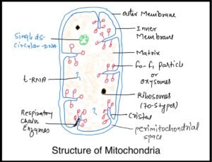
Outer Membrane
The outer membrane is smooth. It is permeable to a number of metabolites.
It is due to presence of protein channels called Porins or which form minute pores. A few enzymes connected with lipid synthesis, are located in the membrane
Inner Membrane
It is permeable to only some metabolites.
It is rich in double phospholipids called cardiolipin (having four fatty acids) which makes the membrane impermeable to ions.
The inner membrane is enfolded (finger like inward folding towards matrix side) variously to form involutions called cristae.
They are meant for increasing the surface area of the inner membrane. Cristae enclose a space that is continuation of the outer chamber.
The density of cristae indicates the intensity of respiration. Inner membrane has the components of respiratory chain enzymes or ETS (electron transport system)
Elementary Particle (F0 – F1 particles or Oxysomes)
The inner membrane as well as its cristae possess small paper pin like structures named elementary particles or F0 – F1 particles or Oxysomes (or Oxysomes).

A mitochondrion contains 1 x 10⁴ – 1 x 10⁵ elementary particles or F0-F1 particles.
Each F0 – F1 particles has a head, a stalk and a base. The base (F0 subunits) is about 11 nm long and 1.5 nm in thickness.
The stalk is 5 nm long and 3.5 nm broad. The head (F1 subunit) has diameter of 8.5 nano meter(nm).
F0 – F1 particles functions as ATP-ase. Hence these are the site of ATP synthesis during Oxidative Phosphorylation.
Enzymes of electron transport are located in the inner membrane in contact with elementary particles.
Adhesion Sites
At places, outer and inner mitochondrial membranes come in contact. They are called adhesion sites.
Adhesion sites are special permeation regions of the mitochondrion for transfer of materials from outside to inside and vice versa.
Outer Chamber (Perimitochondrial Space)
It is present between the outer and inner membrane of the mitochondrial envelope. Usually, it is 60 – 100 angstrom wide.
It extends into the spaces of the cristae. The chamber contains a fluid having a few enzymes.
Inner Chamber
- It forms the core of the mitochondrion. The inner chamber contains a semi – fluid matrix.
- The matrix has
- Protein particles
- 70 S-Ribosomes which is resemble to prokaryotic ribosomes
- RNAs (t-RNA)
- DNA (mitochondrial or mt-DNA)-a single circular double stranded.
- Enzymes of Krebs or TCA cycle (except succinate dehydrogenase which has membrane based location).
- Enzymes of amino acid and fatty acid metabolism
Autonomy of Mitochondria (Semiautonomous Cell Organelles)
- Mitochondria Consider as Semiautonomous cell Organelles due to following Reasons-
- Mitochondria have their own DNA which can replicate independently.
- Mitochondrial DNA produces its own m-RNA, t-RNA and r-RNA.
- The organelles possess their own Ribosomes (70 S type).
- Mitochondria synthesise some of their own structural proteins as well as enzymes.
- However most of the mitochondrial proteins or enzymes are synthesized under instructions from nuclear DNA of cell.
- The organelles synthesise some of the enzymes required for their functioning.
- They grow internally.
- New mitochondria develop by division/binary fission of pre-existing mitochondria.
- However, mitochondria are not fully autonomous.
- Both their structure and functioning are partially controlled by nucleus of the cell and availability of materials from cytoplasm.
- Mitochondria are believed to be Symbionts (Margulis, 1971 & According to Endosymbiont Theory)
- In the Eukaryotic cells which became associated with them quite early in the evolution.
Functions of Mitochondria
- Mitochondria are the site aerobic respiration in which respiratory substrates are completely oxidized to carbon dioxide and water.
- The energy liberated in the process is initially stored in the form ATP.
- ATP used in various energy requiring processes of the cell like muscle contraction, nerve impulse conduction, biosynthesis, membrane transport, cell division, movement, etc.
- Because of the formation of ATP, the mitochondria are called power houses of the cell.
- Mitochondria provide important intermediates for the synthesis of several biochemicals like chlorophyll, cytochromes, pyrimidines, steroids, alkaloids, etc.
- The matrix of inner chamber of the mitochondria has enzymes for the synthesis of fatty acids.
- Synthesis of many amino acids occurs in the mitochondria.
- The first formed amino acids are glutamic acid and aspartic acid.
- Mitochondria may store and release Calcium when required.
- An organism generally receives mitochondria from its mother (maternal inheritance).
- Mitochondria are also involved in the Photorespiratory pathway along with chloroplast and Peroxisome during the Photorespiration or C2 Cycle which is found in C3 plants as a wasteful process.
